
最新消息
[新知分享] 關於腦脊髓液的胞外體(CSF-EVs)之研究現狀解析
腦脊髓液(Cerebrospinal Fluid, CSF)內含的胞外體(Extracellular vesicles, EVs), 可做為中樞神經系統疾病的診斷和監測的生物標誌物並具有巨大的潛力。
然而該領域目前面臨缺乏各研究之間的”可重複性”, 使得結果的一致性幾乎不可能有極高的再現情況, 在此我們為大家提出了CSF-EVs的分離純化考量並總結了迄今為止的關鍵發現, 同時也回顧該領域現今發展的相關問題。

簡介:
雖然CSF不像plasma那樣容易獲得, 但其在尋找神經退行性疾病(neurodegenerative diseases)和中樞神經系統(central nervous system, CNS)癌症的生物標誌物中, 包含監測某些治療方法(例如對愛滋病毒患者)與CNS相關的副作用方面等潛力卻是無價的。EVs是屬於”奈米”等級的尺寸且是被雙層脂質包覆的囊泡, 具有蛋白質、RNA和來自原生細胞的各種小分子。由於這些珍貴的”貨物”, 在研究CSF-EVs中尋找疾病的腦源性生物標誌物方面具有重大的意義。
我們也探討了CSF的組成及其對其EVs分離方法的意義, 也會深入探究CSF-EVs並提出該領域的改進方向。

CSF的组成:
CSF包裹著大腦和脊髓, 可提供機械性的保護並同時促進細胞間通信和維持中樞神經系統的穩態。
CSF的組成與plasma有很大的不同(如下表), 其可溶性蛋白程度非常低[1,2], 而CSF-EVs的濃度也明顯較低[3], 但這可能是一個意想不到的研究優勢。
CSF與身體其他部位是相對分離的, 這減少了混雜其他生物液體中EVs的來源, 使CSF-EVs可占有主導性地位, 雖然由於缺乏良好的標記物, 關於CSF中腦源性EVs比例的知識還很有限, 但有研究已發現CSF中有16.1%的腦特異性蛋白是神經元蛋白, 另外83.9%是膠質蛋白[4]。

從CSF中分離EVs的特別注意事項:
從CSF中分離EVs所面臨的挑戰不同於其他生物液體, 例如若以超速離心法(UC)進行雖然可從plasma中分離出大量的顆粒, 但卻不是非常”純淨”的EVs, 可能會引起EVs的生物功能遭到損傷, UC法已被驗證其不能從CSF中分離出有效的EVs[3,5]。
相比之下, 基於尺寸排除色譜(SEC)的qEV提取管柱(qEV 35 nm系列)已被證明可以有效而無損純淨的從CSF中回收EVs粒子[5]。
另一個需要考慮的因素是樣本的體積量, 對於CSF sample來說其樣本體積量是特別少的, 而許多EVs的分離技術卻會需要相對較大的樣本體積量; 如果用的是UC法, 在樣品體積很小的情況下極可能會造成試管坍塌(tubes collapse)和樣品損耗(sample lost), 這就需要對小體積樣品進行”稀釋” 。
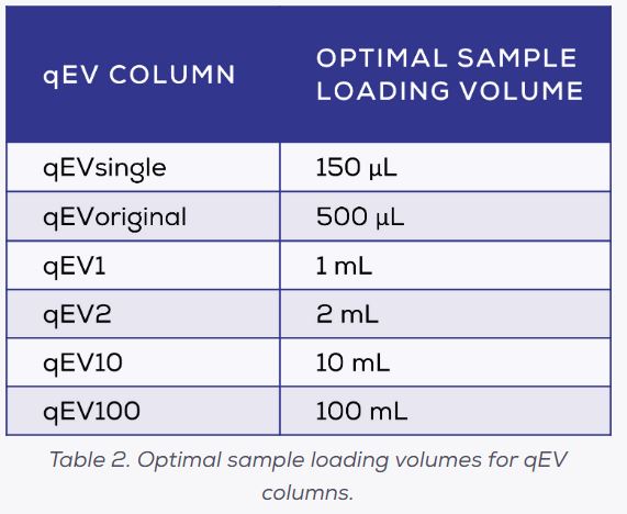
在CSF中EVs的濃度已經是低於其他生物液, 所以在處理過程中儘量減少EVs損失尤為重要, 因此要避免採用預分離中需要做稀釋的技術才是理想的做法, 在此推薦的是qEV column(SEC), 其範圍可適用於幾乎所有的樣品體積: 從150 μL的qEVsingle到最大的100mL的qEV100(如下表)。
另一種分離EVs的方法是使用特定EVs表面蛋白的抗體進行免疫捕獲, 這種方式的純度通常高但產量通常很低, 然而大家都知道EVs在本質上是屬於”異質”的, 其具有不同的表面蛋白質並以不同的程度被攜帶, 因此使用免疫捕獲只能分離出EVs的特定群落。
即使實驗的目標是來自於特定細胞的EVs, 也不能保證來自該細胞的所有EVs都會攜帶特定的標記物, 這意味著其他EVs可能會被遺漏而造成在之後的下游分析中失真。
許多抗體針對的神經元蛋白是L1CAM, 最近的證據也對這種方法提出了質疑並引發了激烈的爭論(詳見我們解析的上一篇), 支持和反對L1CAM作為免疫捕獲的神經元EVs標記的有用性的證據已經被廣泛的討論, 但爭議仍在繼續。
總之雖然免疫捕獲具有一定的特異性, 但其既不是最有效也不是最可靠的技術-SEC法仍然是從CSF中分離純化EVs的較優選擇[5]。

CSF-EVs的醫學重要性:
中樞神經系統疾病一般都非常複雜, 其診斷和監測都具有根本性的困難, 更需要進一步的研究來闡明其發病機制, 這也讓CSF-EVs的研究成為這些領域一種潛在而富有成果的願景。
然而迄今為止的CSF-EVs的研究進度還落後於其他生物液體, 這歸因於其研究並沒有真正的分離出他們的“EVs”而是直接從CSF做分析; 另一些則採用未經驗證的DIY技術(例如在沒有去除可溶性蛋白質的情況下, 使用0.1 μm孔徑過濾[7])。在許多的研究中也沒有遵守MISEV指南, 這可能也說明了CSF-EVs研究通常缺乏一致的結果。

CSF-EVs的主要研究和焦點如上表
包含: 阿茲海默症(Alzheimer’s disease, AD); 帕金森氏症(Parkinson’s disease, PD); 肌萎縮性脊髓側索硬化症(Amyotrophic Lateral Sclerosis, ALS); 多發性硬化(Multiple Sclerosis, MS); 膠質瘤(Glioma)
CSF-EVs未來研究的關鍵問題:
CSF-EVs對於中樞神經系統神經退行性疾病和癌症病理的嚴重程度具有潛在的生物標誌物, 因此對其進行更多的研究會具有重要的醫學意義, 目前該領域仍存在幾個關鍵問題和關注領域, 要能更進一步解決這些問題以提高研究的進程, 並增加辨識有效的生物標誌物和病理機制的可能性:
1. CSF-EVs領域是否有能改進EVs分離純化和表徵的方法?
與其他生物流體中使用的方法相比, 目前該領域內EVs分離方法的品質參差不齊, 因為其具備高度的”異質性”, 意味著不同研究中所分離純化的EVs有著不同的亞群和雜蛋白污染物, 導致研究之間缺乏一致性。
EVs的特徵描述也同樣不夠完善, 許多文獻甚至是最近發表的內容, 都沒有包括任何或只有很少的EVs特徵描述, 建議該領域的研究可以遵循MISEV的EVs分離和表徵標準。
2. CSF-EVs的生物標誌物是否更優於plasma-EVs的生物標誌物?
與plasma sample相比, 因CSF樣品具有較強的侵入性和較少的樣本體積, 故CSF-EVs的生物標誌物具有其獨特的缺陷, 重點是避免對患者進行潛在但不具保證的侵入性手術; 因此若與plama-EVs比較, CSF-EVs的”任何”潛在的生物標誌物都非常的珍貴而重要。
3. CSF儲存的EVs其穩定性如何?
用於生物標誌物研究的CSF在分離EVs之前的儲存條件和保存時間會有很大的差異, 這需要對CSF中儲存EVs的影響進行更全面的研究以確定各種做法對使用這些樣本的研究結果的影響。
參考文獻:
1. Fogh, J. R., Jacobsen, A. M., Nguyen, T., Rand, K. D. & Olsen, L. R. Investigating surrogate cerebrospinal fluid matrix compositions for use in quantitative LC-MS analysis of therapeutic antibodies in the cerebrospinal fluid. Analytical and Bioanalytical Chemistry 412, 1653-1661 (2020). https://doi.org:10.1007/s00216-020-02403-3
2. Hladky, S. B. & Barrand, M. A. Mechanisms of fluid movement into, through and out of the brain: evaluation of the evidence. Fluids Barriers CNS 11, 26 (2014). https://doi.org:10.1186/2045-8118-11-26
3. Skalnikova, H. K. et al. Isolation and Characterization of Small Extracellular Vesicles from Porcine Blood Plasma, Cerebrospinal Fluid, and Seminal Plasma. Proteomes 7 (2019). https://doi.org:10.3390/proteomes7020017
4. Muraoka, S. et al. Proteomic Profiling of Extracellular Vesicles Derived from Cerebrospinal Fluid of Alzheimer's Disease Patients: A Pilot Study. Cells 9 (2020). https://doi.org:10.3390/cells9091959
5. Ter-Ovanesyan, D. et al. Framework for rapid comparison of extracellular vesicle isolation methods. Elife 10 (2021). https://doi.org:10.7554/eLife.70725; 10.7554/eLife.70725.sa1; 10.7554/eLife.70725.sa2
6. Gorgens, A. et al. Identification of storage conditions stabilizing extracellular vesicles preparations. Journal of Extracellular Vesicles 11 (2022). https://doi.org:10.1002/jev2.12238
7. Wang, M. et al. Exosomal LGALS9 in the cerebrospinal fluid of glioblastoma patients suppressed dendritic cell antigen presentation and cytotoxic T-cell immunity. Cell Death & Disease 11 (2020). https://doi.org:10.1038/s41419-020-03042-3
8. Spitzer, P. et al. Microvesicles from cerebrospinal fluid of patients with Alzheimer's disease display reduced concentrations of tau and APP protein. Scientific Reports 9 (2019). https://doi.org:10.1038/s41598-019-43607-7
9. Eitan, E. et al. Extracellular Vesicle-Associated Aβ Mediates Trans-Neuronal Bioenergetic and Ca(2+)-Handling Deficits in Alzheimer's Disease Models. NPJ Aging Mech Dis 2, 16019- (2016). https://doi.org:10.1038/npjamd.2016.19
10. Sandau, U. S. et al. Differential Effects of APOE Genotype on MicroRNA Cargo of Cerebrospinal Fluid Extracellular Vesicles in Females With Alzheimer's Disease Compared to Males. Frontiers in Cell and Developmental Biology 10 (2022). https://doi.org:10.3389/fcell.2022.864022
11. Tan, Y. J. et al. Altered Cerebrospinal Fluid Exosomal microRNA Levels in Young-Onset Alzheimer's Disease and Frontotemporal Dementia. Journal of Alzheimers Disease Reports 5, 805-813 (2021). https://doi.org:10.3233/adr-210311
12. Longobardi, A. et al. Cerebrospinal Fluid EV Concentration and Size Are Altered in Alzheimer's Disease and Dementia with Lewy Bodies. Cells 11 (2022). https://doi.org:10.3390/cells11030462
13. Vacchi, E. et al. Profiling Inflammatory Extracellular Vesicles in Plasma and Cerebrospinal Fluid: An Optimized Diagnostic Model for Parkinson's Disease. Biomedicines 9 (2021). https://doi.org:10.3390/biomedicines9030230
14. Otake, K., Kamiguchi, H. & Hirozane, Y. Identification of biomarkers for amyotrophic lateral sclerosis by comprehensive analysis of exosomal mRNAs in human cerebrospinal fluid. Bmc Medical Genomics 12 (2019). https://doi.org:10.1186/s12920-019-0473-z
15. Thompson, A. G. et al. CSF extracellular vesicle proteomics demonstrates altered protein homeostasis in amyotrophic lateral sclerosis. Clinical Proteomics 17 (2020). https://doi.org:10.1186/s12014-020-09294-7
16. Sjoqvist, S. & Otake, K. A pilot study using proximity extension assay of cerebrospinal fluid and its extracellular vesicles identifies novel amyotrophic lateral sclerosis biomarker candidates. Biochemical and Biophysical Research Communications 613, 166-173 (2022). https://doi.org:10.1016/j.bbrc.2022.04.127
17. Hayashi, N. et al. Proteomic analysis of exosome-enriched fractions derived from cerebrospinal fluid of amyotrophic lateral sclerosis patients. Neuroscience Research 160, 43-49 (2020). https://doi.org:10.1016/j.neures.2019.10.010
18. Welton, J. L. et al. Cerebrospinal fluid extracellular vesicle enrichment for protein biomarker discovery in neurological disease; multiple sclerosis. Journal of Extracellular Vesicles 6 (2017). https://doi.org:10.1080/20013078.2017.1369805
19. Geraci, F. et al. Differences in Intercellular Communication During Clinical Relapse and Gadolinium-Enhanced MRI in Patients With Relapsing Remitting Multiple Sclerosis: A Study of the Composition of Extracellular Vesicles in Cerebrospinal Fluid. Frontiers in Cellular Neuroscience 12 (2018). https://doi.org:10.3389/fncel.2018.00418
20. Verhaak, R. G. W. et al. Integrated Genomic Analysis Identifies Clinically Relevant Subtypes of Glioblastoma Characterized by Abnormalities in PDGFRA, IDH1, EGFR, and NF1. Cancer Cell 17, 98-110 (2010). https://doi.org:10.1016/j.ccr.2009.12.020
21. Figueroa, J. M. et al. Detection of wild-type EGFR amplification and EGFRvIII mutation in CSF-derived extracellular vesicles of glioblastoma patients. Neuro-Oncology 19, 1494-1502 (2017). https://doi.org:10.1093/neuonc/nox085
22. Xu, H. et al. miR-3184-3p enriched in cerebrospinal fluid exosomes contributes to progression of glioma and promotes M2-like macrophage polarization. Cancer Science 113, 2668-2680 (2022). https://doi.org:10.1111/cas.15372
22. Qi, Y. H. et al. The dual role of glioma exosomal microRNAs: glioma eliminates tumor suppressor miR-1298-5p via exosomes to promote immunosuppressive effects of MDSCs. Cell Death & Disease 13 (2022). https://doi.org:10.1038/s41419-022-04872-z